In this article
Fascia has become a familiar word in fitness, often attached to a roller, a treatment, or a new way of moving. Some explanations make it sound like the missing answer to every tight muscle and recurring discomfort. Others dismiss it as marketing. Neither position helps someone decide how to train.
The useful middle ground is straightforward: connective tissue belongs in our understanding of movement, but identifying a tissue does not automatically establish a diagnosis, a treatment, or a superior exercise method.
For an active adult, the practical question is what the body needs to tolerate and perform. Can you produce force through a useful range, manage a landing, rotate under control, and recover well enough to repeat the activity? Fascia contributes to the system doing that work. It does not operate separately from muscles, tendons, joints, and the nervous system.
A connected structure, not a separate training system
The word fascia commonly refers to connective-tissue sheets and layers surrounding, separating, and linking structures. Broader descriptions of the fascial system include additional connective tissues. Definitions vary, which is one reason claims about “fascia training” need to identify what tissue and outcome they actually mean.
These tissues are not inert packaging. Their mechanical relationships help distribute forces, and their nerve supply contributes sensory information. A 2022 review of fascial innervation identified sensory nerve structures across the tissues examined, while also finding differences by anatomical region and study method. The evidence included both animal and human studies. It supports a sensory role; it does not establish that fascia explains an individual person’s pain or balance problem.
This fits the AEON Systems Lens. We consider the relationships among tissues and physical capacities, then choose the training priorities that matter for the person. A connected body does not require an elaborate explanation for every movement difference.
Seeing tissue move is not proof of a treatment
Researchers can investigate fascia during ordinary strength movements. In a 2023 case–control study, ultrasound was used to examine thoracolumbar fascia during deadlifting and trunk extension in athletes, untrained adults, and people with acute low back pain. The study involved 48 participants and found differences between groups and relationships with movement velocity.
That is useful evidence that the tissue participates in movement. Because this was a group comparison rather than a training intervention, it cannot show that a specific exercise repairs fascia, that a measured difference causes pain, or that every person should deadlift. The researchers also identified limitations in load standardization and measurement.
The distinction matters whenever a scientific image becomes a sales claim. Showing that a structure changes during a task is different from demonstrating that a proposed treatment improves an outcome people care about.
A change in flexibility is not the same as new capacity
Foam rolling can have a practical place without requiring a story about breaking adhesions or permanently reshaping tissue. A useful result is still useful when its mechanism remains uncertain.
An eight-week randomized trial in recreationally active adults found that foam rolling improved the stand-and-reach flexibility measure compared with the comparison groups. It did not find significant improvements in the balance and muscle-performance measures tested. The trial was small, and its findings should not be extended to every rolling method or population.
The lesson is about matching the tool to the goal. Feeling less restricted or reaching farther does not demonstrate better strength, faster court movement, or greater tolerance for repeated loading. Nor does it establish that fascia was physically “released.” If a brief rolling routine makes a warm-up more comfortable, it may be worth keeping. It should earn its place through the result, not through an unverified tissue explanation.
Loading evidence needs tissue-specific interpretation
Connective tissues can adapt to training, but findings about one structure should not be applied indiscriminately to every other structure. Tendon research provides a useful example, not a universal fascia prescription.
A systematic review of loading interventions in healthy adults examined Achilles and patellar tendons. It found changes in mechanical properties, with loading magnitude influencing the response. The included participants were 18–50 years old, and interventions lasted at least eight weeks. These results support progressive loading of those tendons; they do not prove that all fascial tissues adapt at the same rate or need the same exercise.
“Stiffness” also has a technical meaning in this research: resistance to deformation under load. It is not interchangeable with the everyday feeling of being stiff. A change in a laboratory property is not automatically good or bad without considering function and context. Our Achilles capacity article develops that distinction for the lower leg.
What this changes in a training session
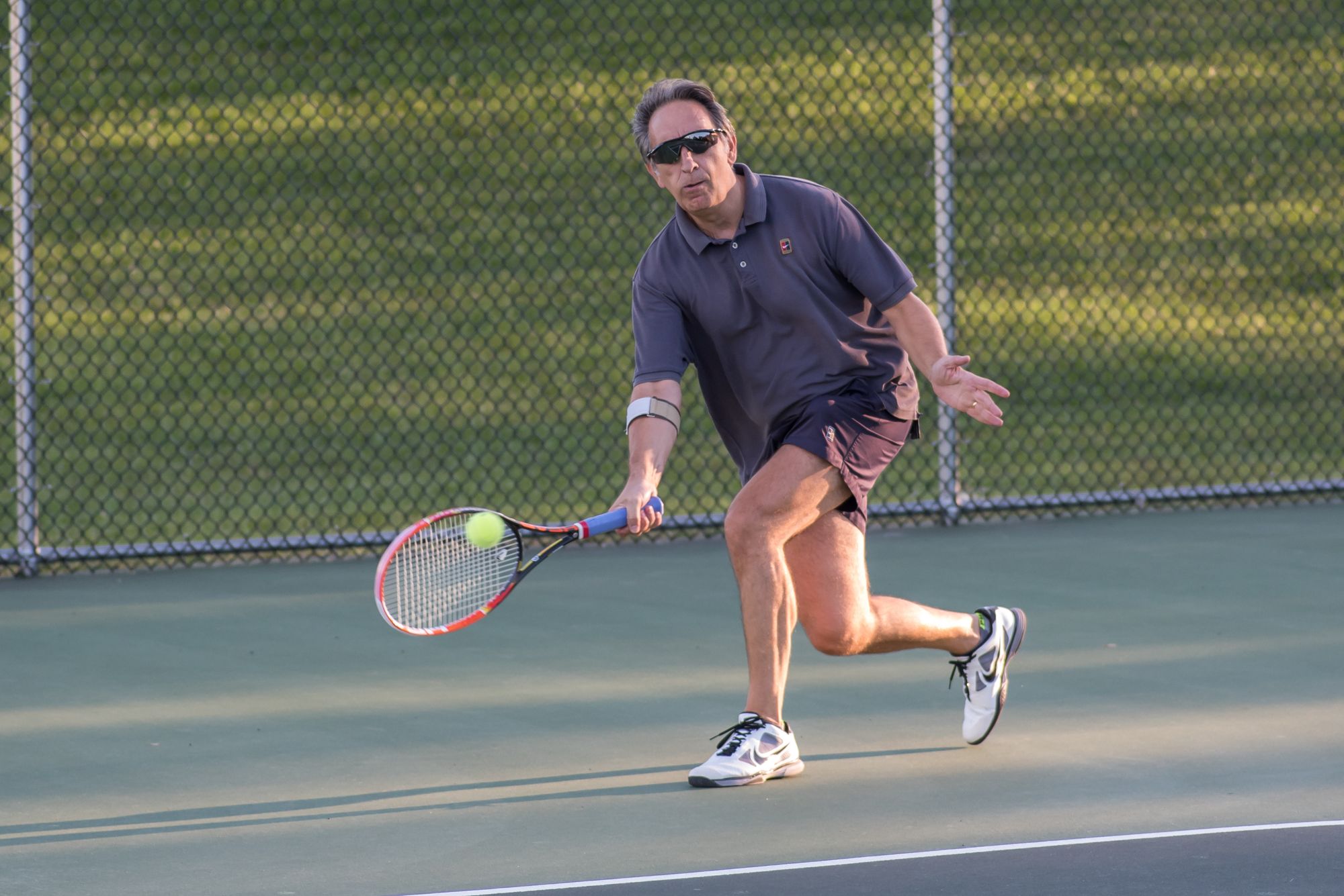
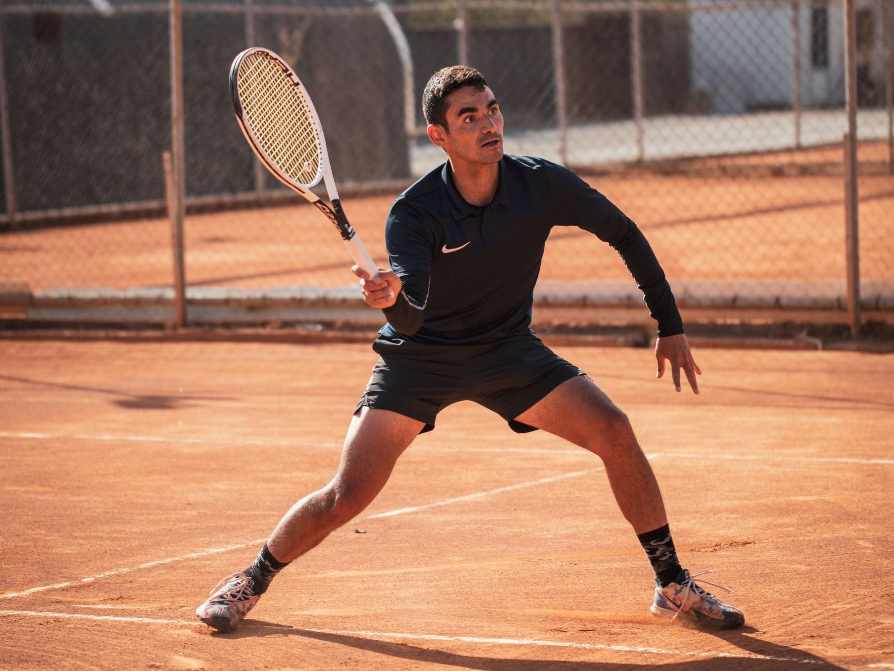
The coaching application is to develop the capacities required by the activity, rather than organize the session around one tissue label. For a recreational tennis player, that could mean the following progression. This is a planning example, not a standardized fascia protocol.
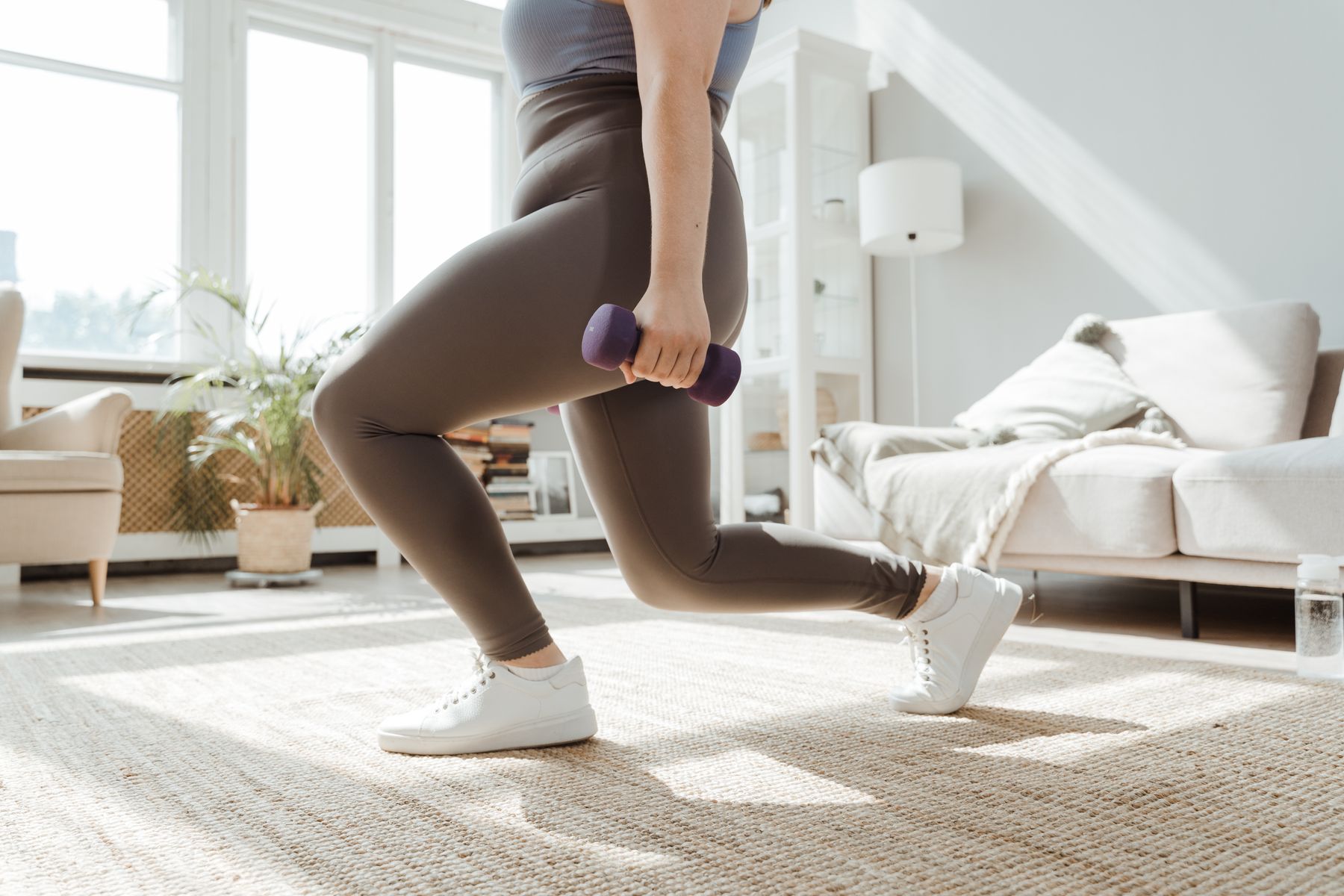
Establish controllable strength. Choose suitable squat, hinge, calf, push, pull, and trunk exercises. Select resistance and range around the person’s current ability and history, not an impressive-looking exercise.
Use more than one movement direction. When relevant, add controlled lateral steps, split stances, reaching, and rotation. These are ways to practice the demands of the task, not evidence that a named fascial chain has been corrected.
Prepare for faster demands separately. Tennis eventually asks for rapid push-off, braking, and repeated changes of direction. Appropriate elastic and reactive work can follow a manageable foundation; stretching and rolling do not substitute for that preparation.
Review the response. Track repeatable movement, useful range, training consistency, recovery, and relevant sport performance. Change one important demand at a time so the effect is easier to interpret.
This is Minimum Effective Complexity: understand the connected system, but prescribe only what the client can use. Biomechanics training should make decisions clearer, not add a new corrective ritual to every session.
Keep observation separate from diagnosis
AEON uses SCREEN / TRAIN / REFER to maintain that boundary. SCREEN means understanding goals, history, symptoms, current activity, and response to movement. It does not mean diagnosing fascial restrictions from posture or touch.
TRAIN means working on modifiable capacity through appropriate strength, mobility, coordination, conditioning, and progression. REFER means recognizing when persistent or worsening pain, neurological symptoms, unexplained functional loss, or another concerning change calls for an appropriately qualified clinician rather than another drill.
Fascia deserves attention because the body is connected. It does not need to become the organizing explanation for everything the body does. The standard for progress remains practical: more usable capability for the activities you want to keep doing.
Selected research
- Fascial Innervation: A Systematic Review of the LiteratureSuarez-Rodriguez et al. International Journal of Molecular Sciences. 2022;23(10):5674.
- Thoracolumbar fascia deformation during deadlifting and trunk extension in individuals with and without back painBrandl et al. Frontiers in Medicine. 2023;10:1177146. Case–control study.
- The Training Effects of Foam Rolling on Core Strength Endurance, Balance, Muscle Performance and Range of Motion: A Randomized Controlled TrialJunker and Stöggl. Journal of Sports Science & Medicine. 2019;18(2):229–238.
- Human tendon adaptation in response to mechanical loading: a systematic review and meta-analysis of exercise intervention studies on healthy adultsBohm, Mersmann and Arampatzis. Sports Medicine – Open. 2015;1:7.